Ultrasound
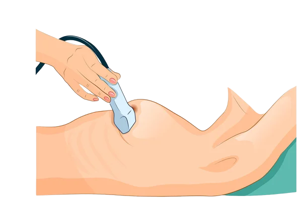
Ultrasounds are highly effective imaging procedures that use sound waves to create detailed images of the inside of your body. Such images provide valuable information your provider uses to diagnose possible medical issues and establish an appropriate treatment plan. The ultrasound technologist completes many ultrasounds by placing a transducer on the outside of your body. However, some ultrasounds require placing a small device inside the body.
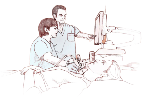
What is an ultrasound?
Ultrasounds are highly effective imaging procedures that use sound waves to create detailed images of the inside of your body. Such images provide valuable information your provider uses to diagnose possible medical issues and establish an appropriate treatment plan. The ultrasound technologist completes many ultrasounds by placing a transducer on the outside of your body. However, some ultrasounds require placing a small device inside the body.
How should I prepare for an ultrasound?
There’s usually no special preparation needed before you undergo an ultrasound, with a few exceptions. In some cases, your specialist asks you to avoid food and drink for a period of time before the ultrasound. Other times drinking water and having a full bladder is beneficial. Wear loose, comfortable clothing or change into a hospital gown if asked to by your provider. Remove any jewelry if necessary. For some ultrasound procedures, you can take a sedative to help you feel more relaxed.
What happens during an ultrasound?
During an ultrasound, you lie on an exam table or rest in a comfortable, reclining chair. Your specialist applies a gel to the areas being examined and slides a transducer on the outside or inside of your body to create sound-wave images. As sound waves bounce off specific areas of your body, they send and capture images on a computer screen. The time it takes to complete an ultrasound varies depending on the type you undergo, but it usually lasts about 30-60 minutes. You can often return to typical daily activities immediately.
What should I expect after the procedure?
After you complete an ultrasound, your specialist reviews the results with you and determines which treatment is most appropriate. They may suggest rest, watchful waiting, additional diagnostic testing, medications, minor procedures, or care from another specialist.
To schedule an appointment with SonoStat Imaging you must contact your physician who will determine if you’re a candidate for an on-site ultrasound. Also, you may call SonoStat Imaging and our team will schedule you ultrasound at our nearest location in your area.
What does an ultrasound diagnose?
SonoStat Imaging offers ultrasound Imaging at the physicians offices if you have symptoms or risk factors of:
* Pregnancy
* Gynecological conditions
* Gallbladder diseases
* Blood flow irregularities
* Breast lumps
* Thyroid gland issues
* Prostate or genital problems
* Tumors
* Metabolic bone diseases
* Joint inflammation
* Heart or any cardiovascular disease
An ultrasound can also guide needle placement during a biopsy or tumor treatment. Your specialist reviews your symptoms, medical history, lifestyle, and more. They also check your vital signs and complete a physical exam to determine if you’re a candidate for an ultrasound.
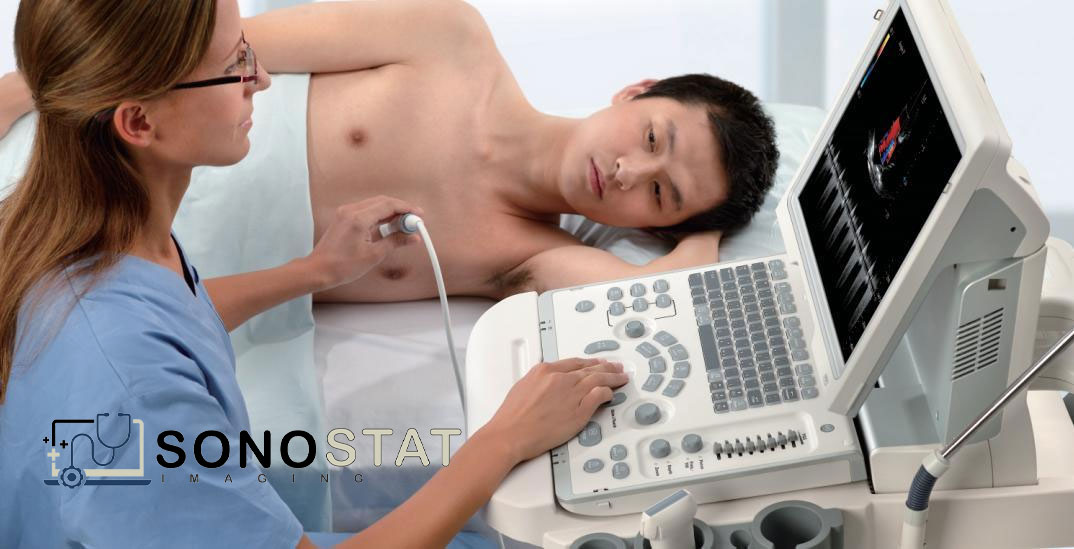

Therapeutic Ultrasounds
Therapeutic ultrasounds are used to modify or destroy tissue, such as blood clots, kidney stones or tumors using a process called thermal ablation. The benefit of therapeutic ultrasound as a non-invasive therapy is still being studied, though patients generally prefer the lack of incisions and recovery time when compared to traditional surgery. Unlike diagnostic ultrasounds, therapeutic ultrasounds are only performed as interventional radiology procedures in one of our hospitals
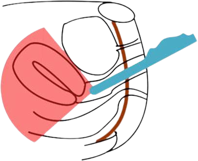
Pelvic ultrasounds
Pelvic ultrasounds are focused on the structures and organs of the lower abdomen and pelvis. There are two types of pelvic ultrasound: transabdominal and transvaginal (for women). These exams are frequently used to evaluate the reproductive and urinary systems
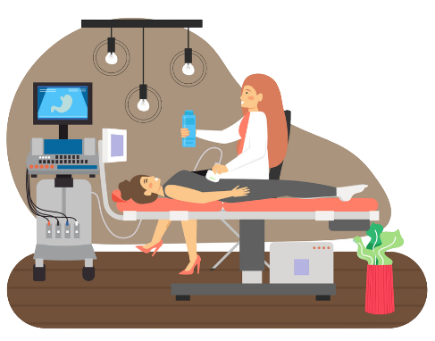
Abdominal ultrasounds
Abdominal ultrasounds scan the upper abdominal area. They are often used to help diagnose pain or discomfort of the liver, gallbladder, bile ducts, pancreas, kidneys, spleen and abdominal aorta.

Obstetric ultrasounds
Obstetric ultrasounds produce sonogram images of an embryo or fetus within a pregnant woman, as well as the mother’s uterus and ovaries. These ultrasounds, which do not use radiation, are the preferred imaging method for monitoring pregnant women and their unborn babies. Doppler ultrasounds, which evaluate blood flow in the umbilical cord, fetus or placenta, may also be part of this exam.

Musculoskeletal ultrasounds
Musculoskeletal ultrasounds evaluate muscles, tendons, ligaments and joints throughout the body. This procedure can help your physician diagnose sprains, strains, tears and other soft tissue conditions, as well as, evaluate your need for physical or occupational therapy. Musculoskeletal ultrasounds are sometimes used to help physicians conduct guided steroidal injections for managing inflammation
Breast ultrasounds
Uses high-frequency sound waves to create ultrasound images of the hemodialysis access site. Doctors use this exam to evaluate the rate of blood flow through the arteries and to detect any blot clots or blockages in the vessels. We perform specific duplex exams to assess maturity, patency and function of arteriovenous dialysis fistulas and grafts, which are surgically created to improve long-term vascular access for hemodialysis patients
Abdominal and aortic duplex exam
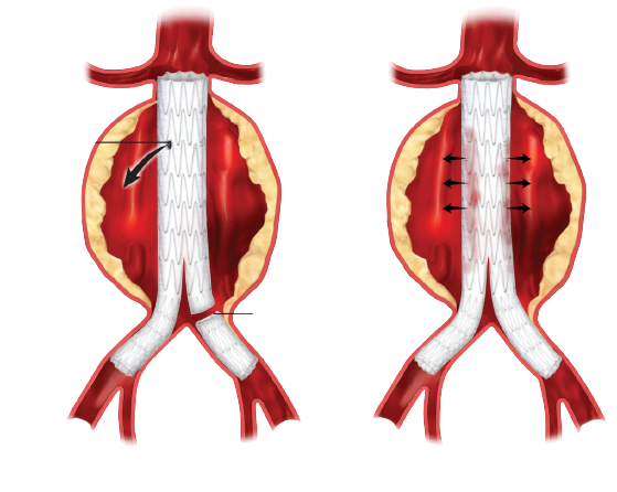
Uses high-frequency sound waves to create ultrasound images of the abdominal aorta. Doctors use this exam to evaluate blood flow through the aorta toward the legs, and to measure the aorta’s diameter to detect and monitor for potentially life-threatening aortic aneurysm. Doctors also perform this exam after aortic endograft repair, as well as to study the renal arteries and mesenteric arteries, which supply blood to the large and small intestines
