Our Services
We are partnered with several recognized Radiology facilities and offer services for MRI, CT, Sedation MRI, Coronary Calcium scan and X-Ray. We are in-Network for most insurances and also offer competitive cash prices for all radiology services
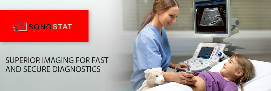
We are a mobile diagnostic Imaging company offering services in Cardiac, General and Vascular Ultrasound in the DFW Metroplex. Our Ultrasound technologist use the latest technology in ultrasound imaging, including Doppler ultrasounds, to visualize blood flow. Ultrasound use sound waves that bounce off surfaces in the body to produce images of soft tissues. Our services include:
Transthoracic Echocardiography (TTE) 2D, M-Mode (Adult)
The format of 2D echocardiography is well suited to analyze congenital heart disease, consequences of coronary artery disease, and distortions of anatomy due to acquired heart disease
Stress Echocardiogram (Adult)
A stress echocardiogram is a test done to assess how well the heart works under stress. The “stress” can be triggered by either exercise on a treadmill or a medicine called dobutamine. A dobutamine stress echocardiogram (DSE) may be used if you are unable to exercise

Carotid Artery Color Doppler
A carotid artery Doppler ultrasound is a diagnostic test used to check the circulation in the large arteries in the neck. This exam shows any blockage in the veins by a blood clot or “thrombus” formation
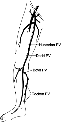
Venous duplex exam
Uses high-frequency sound waves to produce images of veins in the body. Doctors use the venous duplex exam to examine for blood clots or deep vein thrombosis (DVT), which is potentially life-threatening. We also use this technique to evaluate for venous reflux or insufficiency, a condition that develops when valves inside of veins become weakened or damaged. This causes blood-flow reversal and pooling of blood in the lower extremity veins, especially when standing
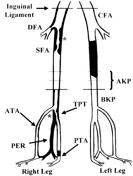
Arterial duplex exam
Uses high-frequency sound waves to produce images of major arteries in the arms and legs. Doctors use this exam to evaluate blood flow through the arteries and to detect any blockages or narrowing in the vessels. Your doctor may also recommend these studies to detect the presence, severity and location of arterial diseases
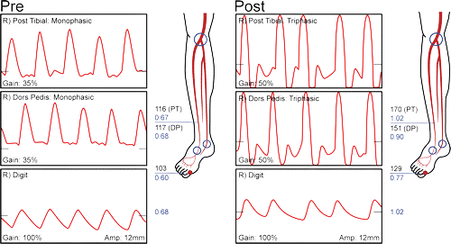
Arterial physiologic Doppler exam
Uses high-frequency sound waves to estimate blood flow through the arteries in the arms and legs. This test relies on a device that emits high-frequency sound waves as well as blood pressure cuffs to evaluate for blockages. It records blood-flow patterns and compares blood pressures at various levels of the arms and legs.

Thyroid Ultrasound
Thyroid ultrasound is a sound wave picture of the thyroid gland taken by a hand-held instrument and translated to a 2-dimensional picture on a monitor. It is used in diagnosis of tumors, cysts or goiters of the thyroid, and is a painless, no-risk procedure.
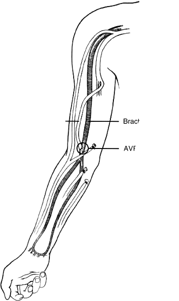
Duplex of the hemodialysis access (arteriovenous fistula or grafts)
Uses high-frequency sound waves to create ultrasound images of the hemodialysis access site. Doctors use this exam to evaluate the rate of blood flow through the arteries and to detect any blot clots or blockages in the vessels. We perform specific duplex exams to assess maturity, patency and function of arteriovenous dialysis fistulas and grafts, which are surgically created to improve long-term vascular access for hemodialysis patients
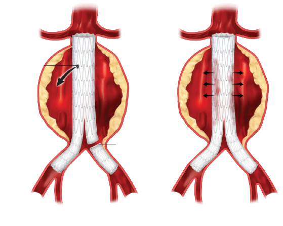
Abdominal and aortic duplex exam
Uses high-frequency sound waves to create ultrasound images of the abdominal aorta. Doctors use this exam to evaluate blood flow through the aorta toward the legs, and to measure the aorta’s diameter to detect and monitor for potentially life-threatening aortic aneurysm. Doctors also perform this exam after aortic endograft repair, as well as to study the renal arteries and mesenteric arteries, which supply blood to the large and small intestines

Thyroid Ultrasound
Thyroid ultrasound is a sound wave picture of the thyroid gland taken by a hand-held instrument and translated to a 2-dimensional picture on a monitor. It is used in diagnosis of tumors, cysts or goiters of the thyroid, and is a painless, no-risk procedure.
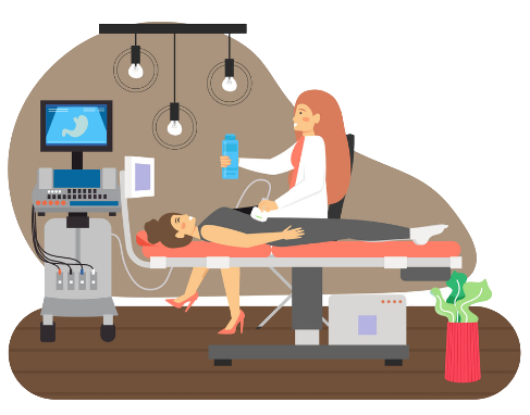
Abdominal ultrasounds
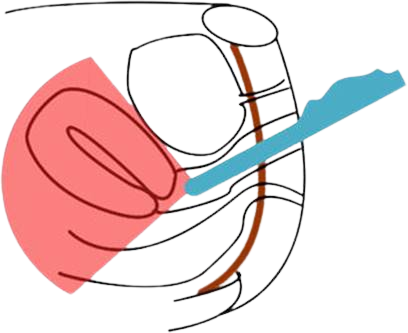
Pelvic ultrasounds
Pelvic ultrasounds are focused on the structures and organs of the lower abdomen and pelvis. There are two types of pelvic ultrasound: transabdominal and transvaginal (for women). These exams are frequently used to evaluate the reproductive and urinary systems
Say Goodbye to Health Woes & Hello to Optimal Wellness
What to Expect
Mobile services available 24/7
- Ultrasound
- Abdominal ultrasonography
- Transvaginal ultrasound
- Obstetric ultrasonography
- 3D ultrasound
Advance Ultrasound 27/7
- Echocardiogram
- Transrectal ultrasonography
- Abdomen
- Doppler ultrasonography
- Pelvic Ultrasound
